Brian Saltzman MD. Beth Israel-Deaconess Medical Center Harvard Medical School, Boston, MA. and Josef Hochman, BSc. EE.Direx Systems Corporation, Natick, MA
American Urology Associaton Congress
Focal Cross-Section, the Truncated Focal Area and the Truncated Volume:
Abstract
The Peak Pressure at F2 and the Focal Area are the traditional parameters used to compare the performance and effectiveness of the Shock Wave produced by different lithotripters.
Lately, new electromagnetic lithotripters were introduced, some with higher Focal Peak Pressure. This fact may lead to believe that they are more efficient than the traditional spark gap systems.
At the same time all electromagnetic systems have very thin focal areas, much smaller than the typical stone size, and therefore the available energy is not optimized for stone fragmentation, usually requiring much more shocks compared to a traditional Electrohydraulic lithotripter.
The Focal Cross Section at F2, the Truncated Focal Area and Volume are 3 new tools which allow a more accurate evaluation of the Shock Wave characteristics and efficiency of different lithotripters.
Eleven currently used lithotripters including the Dornier HM-3 were compared:
The results show two categories of Lithotripters:
a) Large Focus: Dornier HM-3, Medstone STS-T, Direx Tripter Compact and Medispec Econolith.
b) Small Focus: All electromagnetic lithotripters, plus Edap Praktis and the Healthronics Lithotron.
The Average Focal Cross Section for Large Focus lithotripters is 5 times bigger than the small ones.
The Average Truncated Area is 2.35 times bigger and the Average Truncated Volume is 5 times bigger in Big Focus Lithotripters compared to Small Focus ones.
This may help to explain why usually the electromagnetic lithotripters require much more shocks to break stones and have larger retreatment rates.
Introduction
Various lithotripters using different Shock Wave technologies are currently offered to treat stones in the urinary tract.
In order to compare the various systems offered, Urologists analyze their technical specifications to evaluate their performance. (Ref 1)
Traditionally the Peak Bar Pressure at F2 is the first parameter considered as an indicator of the available energy of a lithotripter and, therefore, has served as a first indicator of the efficiency of the system.
Some confusion existed in the past regarding the numerical value of this Pressure at F2.
Due to specific conditions of the Shock Waves, the measurements done with the older Piezoelectric Crystal sensors lead to erroneous high values of pressure (above thousand bars).
During the last years a new precise sensor made of a membrane of Poly Vinyl Duo Fluoride (PVDF) was developed and adopted by FDA as the only one to be used in Pressure measurements (Ref 2).
Using PVDF, the pressure values recorded are "smaller" compared to old Piezoelectric Crystal sensors, but obviously this is are more accurate and "real" values.
Recently, new Electromagnetic lithotripters were introduced some of them with higher Peak Bar Pressure. This fact leads one to believe that they are more efficient than the traditional spark gap systems.
Looking carefully we can see that this may be misleading.
A correct analysis requires one to look not only at the Peak Bar pressure but also at the focal area geometric dimensions.
All Electromagnetic systems have very thin focal areas - much smaller than the typical stone size-and, therefore the available energy is not optimized for stone fragmentation.
The Total Focal Area which is also used sometimes to compare different lithotripters may be misleading too, because it does not take into account the fact that the typical stone size is much smaller than the long axis of the Focal ellipse and therefore a big portion of the energy is not applied to the stone.
In order to clarify this issue three new tools were developed and are presented:
a) The Focal Cross Section
b) The Truncated Ellipse Focal Area
c) The Truncated Focal Volume.
They will allow a more precise geometrical comparison of the Focal Areas of different lithotripters and hence their effectiveness.
Materials and Methods
Specifications of 11 currently used lithotripters were used from published references (Ref 1).
The distribution of pressure of a lithotripter is centered at the focal point F2 and includes all points whose pressure is between 100% ( f2) and 50% of the Peak Power( 6 dB).
The shape of this focal volume is approximately an ellipsoid (a "cigar" or "watermelon" shape) (Ref 2).
This ellipsoid volume is obtained by rotating the focal ellipse around the long axis.
The geometric specifications of the focal area are the Long Dimension (LD) and Short Dimension (SD) of the ellipse and ellipsoid. The (a) Long radius and (b) Short Radius equal half of the previous values respectively.
1) Focal Area Cross Section (FACS)
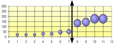
The easiest way of visualizing how much of the stone is subjected to pressure is to look at the Cross Section of the ellipsoid at F2 (Like "cutting" the ellipsoid/cigar at F2 and looking at the circle that originated).
We can calculate the Cross Section area using the formula of the circle area
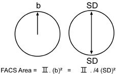 |
| b = Focal Short Radius =SD/2 SD=Focal Short Dimension (diameter) |
2) Ellipse shape geometric function
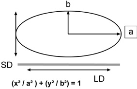
3) Ellipse Focal Area (EFA)
Using the ellipse Long Radius a , and the Short Radius b, we can calculate the full Ellipse area using the formula
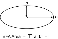
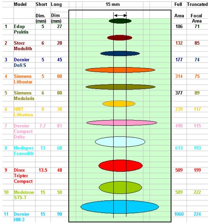
4) Truncated Ellipse Focal Area (TEFA)
Using the Long Radius a, and the Short Radius b, we can calculate the Truncated Ellipse area using the formula5) Ellipsoid Focal Volume (EFV)
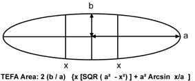
5) Ellipsoid Focal Volume (EFV)
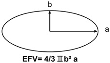
6) Truncated Ellipsoid Focal Volume (TEFV)
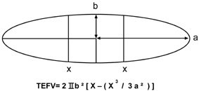
Results
Calculations and graphs were made using the Excel (Microsoft) Program 1)
1) Focal Area Cross Section
| Focal Cross Sections at F2 | ||||
| Circle # | Manufacturer | Model | Diameter | Cross Section |
| Short Dimension | Area | |||
| SD(mm) | (mm 2) | |||
| 1 | 1 Dornier | Doli S | 5 | 20 |
| 2 | Siemens | Lithostar | 5 | 20 |
| 3 | Edap | Praktis | 5 | 20 |
| 4 | Storz | Modulith | 6 | 28 |
| 5 | Siemens | Modularis | 6 | 28 |
| 6 | Dornier | Compact Delta | 7.7 | 47 |
| 7 | Healthronics | Lithotron | 8 | 50 |
| 8 | Medispec | Econolith | 13 | 133 |
| 9 | Direx | Tripter Compact | 13.5 | 143 |
| 10 | Medstone | STS-T | 15 | 177 |
| 11 | Dornier | HM-3 | 15 | 177 |
| Average Large Focal Areas Standard Deviation Average Small Focal Areas Standard Deviation | 157 | |||
| 23 | ||||
| 30 | ||||
| 13 | ||||
 | |
| Small Focal Areas | Large Focal Areas |
| Table and Graph # 1 | |
2) The Truncated Ellipse Focal Area
The Graph below represents all focal ellipses and their truncation.
| Truncation of Focal Ellipses |
 |
| Table and Graph # 2 |
3) Truncated Ellipse Focal Area
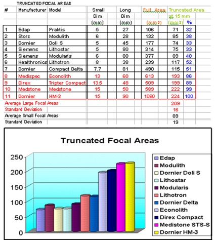 |
| Table and Graph # 3 |
4) Truncated Ellipsoid Focal Volume
 |
| Table and Graph #4 |
Discussion
Analyzing the results shown in Tables #1 through #4, we may distinguish two categories of Lithotripters:
- Large Focus: 4 lithotripters are in this category:
Dornier HM-3, Medstone STS-T, Direx Tripter Compact, and Medispec Econolith.
- Small Focus: 7 Lithotripters are in this category:Storz Modulith, Dornier Doli S, Dornier Compact Delta, Siemens Lithostar, Siemens Modularis (All electromagnetic lithotripters), Edap Praktis, and the Healthronics Lithotron (Spark Gap)
| Cross Section | Cross Section | Truncated Area | Truncated Area | Truncated Volume | Truncated Volume | |
| Average | Standard Deviation | Average | Standard Deviation | Average | Standard Deviation | |
| a)Large Focus | 157 | 23 (15%) | 209 | 16 (8%) | 2306 | 343 (15%) |
| b) Small Focus | 30 | 13 (43%) | 89 | 19 (21%) | 435 | 191 (44%) |
| Ratio a/b | 5.23 | 2.35 | 5.3 |
- The 2 categories of Lithotripters are clearly differentiated, the ration of their Cross Sections, Areas and Volumes are between 2.35 and 5.3.
- The Large Focus group is more homogeneous (Standard Deviation 8 to 15 %) , whereas the Small Focus is less (Standard Deviation 21% to 44 %). This is due to the fact that the Dornier Delta and Healthronics Lithotron have relatively bigger dimensions than the rest of the group, but still far form the Large Focus group.
- ALL Large Focus Lithotripters use the Spark Gap technology.
- ALL Electromagnetic units fall into the Small Focus category.
- Two Spark Gap units are also in the Small Focus category: Edap Praktis and Healthronics Lithotron.
The Edap Praktis, although basically a Spark Device, uses a variation of what is called the Electroconductive Technology.
The purpose of this technology is to reduce the pressure fluctuation between shocks. In order to achieve this, the system uses a special electrode in a highly conductive liquid, with a very small gap and as a result, the focal volume is much smaller than conventional Spark Gap devices.
It can be seen on Graph # 1, that the Large Focus Lithotripters will " cover" most of the stone areas at F2 ( diameter 13 to 15 mm) whereas the Small Focus ones will cover only a fraction of the typical stone.
This may explain why the electromagnetic devices typically require significantly more shocks to adequately fragment kidney stones and also may result in higher retreatment rates.
Recently, concerns have been raised ( Ref 5) regarding the fact that some new Electromagnetic Lithotripters that have very small focal areas and extremely high peak positive pressures are reporting higher clinically significant hematoma rates of 3 to 12% (Ref 6,7 and 8). A trend that is worrisome.
It is becoming clear that the electromagnetic devices with very long and thin focal area/volumes are not suited to fragment stones.
The Truncated Areas and Volumes are intended to advance the discussion relative to the effectiveness of various lithotripters.
References
1. J. Stuart Wolf, Jr. M.D. Issues in choosing a Lithotriptor: Concepts in Design and use. AUA, 2001.
2. Lewin P.A. and Schafer M.E. "Shock Wave sensors: Requirements and Design. J. Lithotripsy and Stone Disease vol. 3 pp 3-17, 1991.
3. IEC International Standard pressure Pulse Lithotripters-Characteristics of Fields. 1998 -04 Annex C, page 21.
4. FDA Guidance for the Content of Premarket Notifications (510 k) for Extracorporeal Shock Wave Lithotripters Indicated for the Fragmentation of Kidney and Ureteral Calculi. August 9, 2000. Page 6.
5. 1st International Consultation on Stone Disease Committee 8: Bioeffects and Physical Mechanisms of SW Effects in SWL. Chairman: James E. Lingeman, M.D. et al.
6. Kohrmann KU, Rassweiler JJ, Manning M, et al. The clinical introduction of a third generation lithotriptor Modulith SL 20. Journal of Urology, 1995; 153:1379-1383.
7. Stefan T, Thorsten B, Chaussy C. Reduced retreatment rate by anatomy related shockwave (SW) energy. Journal of Urology, 1998; 159:S34 (abstract).
8. Piper NY, Dalrymple N, Bishoff JT. Incidence of renal hematoma formation after ESWL using the new Dornier Doli-S lithotriptor. Journal of Urology, 2001; 165:S377 (abstract).

